About this deal
An MRI scanner uses magnetic fields and radio waves to generate images of the inside of the body. Unlike X-rays, an MRI scan can visualise soft tissue such as the organs and blood vessels. It is a safe and painless procedure, leaving no lasting effect on the patient. Because MRI can construct images of soft tissue, it's especially useful for diagnosing joint abnormalities, diseases of the liver and abdominal organs, and identifying tumours and uterine conditions such as fibroids. Yet I was left with more questions than answers. Are these methods used in real-life diagnosis? If not, why not? Why had these women been allowed to live in such pain for so long? More interrogation into the whys and wherefores would have been appreciated.
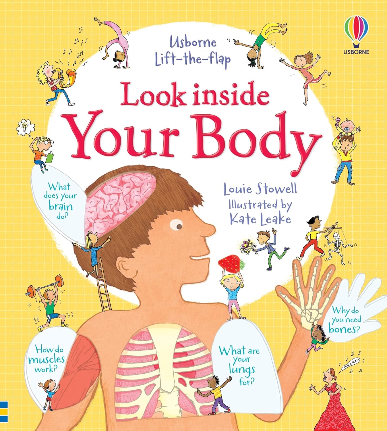
Look Inside Your Body ~ A Best-Selling Body Book! - Surprise Look Inside Your Body ~ A Best-Selling Body Book! - Surprise
When a body is placed between an X-ray source and a photographic (or fluoroscopic) film or screen, an image forms. Denser body parts, such as bones, absorb more X-rays, creating lighter areas on the image. Softer tissue allows X-rays to pass through, leaving dark shadows on the image. W F Bynum and R Porter (eds), Companion Encyclopedia of the History of Medicine (London: Routledge, 1993) Hip bone anyone? ‘Your Body Uncovered with Kate Garraway’ reveals our bodily secrets. (Image credit: BBC) Young readers' minds will boggle as they learn about how their brains work, what happens when they eat, how their lungs use oxygen and much more.Polaroid photograph of ultrasound scan of foetus in utero, taken at University College Hospital, London, 1981 With the help of GPs and surgeons, the show uses 3D imaging technology to show patients exactly what is going wrong in their bodies. From brains and blood to senses and skin - children will love exploring the ins-and-outs of the human body with this fantastic interactive book. In 1973, American chemist Paul Lauterbur (1929–2007) showed that NMR could produce images. British scientist Peter Mansfield (1933–2017) developed the mathematical processes that turned MRI into a useful rapid imaging technique. Lauterbur and Mansfield were awarded the Nobel Prize in Medicine in 2003. J Bronzino, V Smith and M Wade (eds), Medical technology and society: an interdisciplinary perspective (Massachusetts: MIT press 1990)
Look Inside Your Body | Usborne | Be Curious
Ultrasound is a diagnostic imaging technology that uses high-frequency sound waves—well beyond the range of human hearing—to produce pictures of the inside of the body. The technology was impressive, particularly when they compared the size of Hilda’s uterus to a normal one. Gynaecologist Mr Stephen Quinn told her it would be wise to operate, though warned that if there was too much blood loss, he might have to remove her uterus entirely. Despite wanting to have children, Hilda recognised the importance of having the operation and Mr Quinn ended up removing an incredible 100 fibroids. Dr Dimitri Amiras, Trudi and Kate Garraway looking at a GFX representation of Trudi’s frozen shoulder (Photo: BBC/Remarkable TV) Having a serious health condition is terrifying,” says Kate, 54. “When my husband Derek was diagnosed with Covid-19 that fear was made even harder to bear because I didn’t understand what was going on inside his body. Making this series has been an absolutely fascinating process. The incredible augmented reality technology has allowed our contributors to get the most mind-blowing medical consultations, opening their eyes, and mine, to what is going on inside their bodies." A long, thin tube with a small camera inside, called an endoscope, is passed into your body through a natural opening such as your mouth. X-rays were the first technology that made it possible to see inside the body without having to open it up. They were discovered by German physicist Wilhelm Roentgen (1845–1923) at the end of the 1800s and had an immediate impact on anatomical study and diagnostics.
Types of endoscopy
Ultrasound scanners were not commonly used in hospitals until the 1970s. By the 1980s the technology had advanced enough to produce moving images in shades of grey, followed by 3D imaging not long after. Today ultrasound is widely used in surgical procedures and the field of gynaecology. It would be useful throughout KS1-2. Even if the younger children are not fully aware of all the terms they are still able to use their fine motor skills when using the flaps etc. Digital photography continues to play a role in medicine through documentation, research and education. Video cameras are commonly used to look inside the body, most often in the form of endoscopes.
Your Body (Usborne Beginners) : Turnbull, Stephanie, Larkum Your Body (Usborne Beginners) : Turnbull, Stephanie, Larkum
The Challenge season 39: release date, trailer, cast and everything we know about the competition series Scanning technologies collect readings from the body and use a computer to process the data into visual images. The readings can be taken from a variety of sources:
The result watered down an otherwise interesting and innovative programme. It could have easily been a half an hour shorter. It is written in an informative, factual but informal way which is beneficial because it is adding to the children’s vocabulary as well as understanding things that are going on with their own bodies in a fun way.
 Great Deal